Use of preoperative cone-beam computed tomography to aid in establishment of endodontic working length: A systematic review and meta-analysis
| dc.contributor.author | Paterson, A | |
| dc.contributor.author | Franco, V | |
| dc.contributor.author | Patel, S | |
| dc.contributor.author | Foschi, F | |
| dc.date.accessioned | 2023-05-05T12:05:33Z | |
| dc.date.available | 2023-05-05T12:05:33Z | |
| dc.date.issued | 2020 | |
| dc.identifier.issn | 2233-7822 | |
| dc.identifier.issn | 2233-7830 | |
| dc.identifier.uri | https://pearl.plymouth.ac.uk/handle/10026.1/20830 | |
| dc.description.abstract |
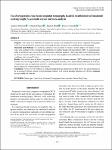
Purpose This study was performed to assess the accuracy of preoperative cone-beam computed tomography (CBCT), when justified for other reasons, in locating the apical foramen and establishing the working length. Materials and Methods Six electronic databases were searched for studies on this subject. All studies, of any type, were included if they compared measurements of working length with preoperative CBCT to measurements using an electronic apex locator (EAL) or histological reference standard. Due to the high levels of heterogeneity, an inverse-variance random-effects model was chosen, and weighted mean differences were obtained with 95% confidence intervals and P values. Results Nine studies were included. Compared to a histological reference standard, CBCT indicated that the apical foramen was on average 0.40 mm coronal of its histological position, with a mean absolute difference of 0.48 mm. Comparisons were also performed to an EAL reference standard, but the conclusions could not be considered robust due to high levels of heterogeneity in the results. Conclusion A low level of evidence is produced suggesting that preoperative CBCT shows the apical foramen to be on average 0.40 mm coronal to its histological position, with a mean absolute difference of 0.48 mm. | |
| dc.format.extent | 183-192 | |
| dc.format.medium | Print-Electronic | |
| dc.language | en | |
| dc.publisher | Korean Academy of Oral and Maxillofacial Radiology | |
| dc.subject | Tooth Apex | |
| dc.subject | Cone-Beam Computed Tomography | |
| dc.subject | Meta-Analysis | |
| dc.subject | Dental Pulp Cavity | |
| dc.title | Use of preoperative cone-beam computed tomography to aid in establishment of endodontic working length: A systematic review and meta-analysis | |
| dc.type | journal-article | |
| dc.type | Review | |
| plymouth.author-url | https://www.ncbi.nlm.nih.gov/pubmed/33005575 | |
| plymouth.issue | 3 | |
| plymouth.volume | 50 | |
| plymouth.publication-status | Published | |
| plymouth.journal | Imaging Science in Dentistry | |
| dc.identifier.doi | 10.5624/isd.2020.50.3.183 | |
| plymouth.organisational-group | |Plymouth | |
| plymouth.organisational-group | |Plymouth|Faculty of Health | |
| plymouth.organisational-group | |Plymouth|Users by role | |
| plymouth.organisational-group | |Plymouth|Users by role|Academics | |
| plymouth.organisational-group | |Plymouth|Faculty of Health|Peninsula Dental School | |
| dc.publisher.place | Korea (South) | |
| dcterms.dateAccepted | 2020-04-26 | |
| dc.date.updated | 2023-05-05T12:05:15Z | |
| dc.rights.embargodate | 2023-8-9 | |
| dc.identifier.eissn | 2233-7830 | |
| dc.rights.embargoperiod | forever | |
| rioxxterms.versionofrecord | 10.5624/isd.2020.50.3.183 |