
Biomarkers for differentiating grade II meningiomas from grade I: a systematic review
| dc.contributor.author | Sofela, AA | |
| dc.contributor.author | McGavin, L | |
| dc.contributor.author | Whitfield, Peter | |
| dc.contributor.author | Hanemann, Clemens Oliver | |
| dc.date.accessioned | 2021-11-11T21:11:05Z | |
| dc.date.available | 2021-11-11T21:11:05Z | |
| dc.date.issued | 2021-06-21 | |
| dc.identifier.issn | 0268-8697 | |
| dc.identifier.issn | 1360-046X | |
| dc.identifier.uri | http://hdl.handle.net/10026.1/18331 | |
| dc.description.abstract |
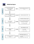
INTRODUCTION: There are a number of prognostic markers (methylation, CDKN2A/B) described to be useful for the stratification of meningiomas. However, there are currently no clinically validated biomarkers for the preoperative prediction of meningioma grade, which is determined by the histological analysis of tissue obtained from surgery. Accurate preoperative biomarkers would inform the pre-surgical assessment of these tumours, their grade and prognosis and refine the decision-making process for treatment. This review is focused on the more controversial grade II tumours, where debate still surrounds the need for adjuvant therapy, repeat surgery and frequency of follow up. METHODS: We evaluated current literature for potential grade II meningioma clinical biomarkers, focusing on radiological, biochemical (blood assays) and immunohistochemical markers for diagnosis and prognosis, and how they can be used to differentiate them from grade I meningiomas using the post-2016 WHO classification. To do this, we conducted a PUBMED, SCOPUS, OVID SP, SciELO, and INFORMA search using the keywords; 'biomarker', 'diagnosis', 'atypical', 'meningioma', 'prognosis', 'grade I', 'grade 1', 'grade II' and 'grade 2'. RESULTS: We identified 1779 papers, 20 of which were eligible for systematic review according to the defined inclusion and exclusion criteria. From the review, we identified radiological characteristics (irregular tumour shape, tumour growth rate faster than 3cm3/year, high peri-tumoural blood flow), blood markers (low serum TIMP1/2, high serum HER2, high plasma Fibulin-2) and histological markers (low H3K27me3, low SMARCE1, low AKAP12, high ARIDB4) that may aid in differentiating grade II from grade I meningiomas. CONCLUSION: Being able to predict meningioma grade at presentation using the radiological and blood markers described may influence management as the likely grade II tumours will be followed up or treated more aggressively, while the histological markers may prognosticate progression or post-treatment recurrence. This to an extent offers a more personalised treatment approach for patients. | |
| dc.format.extent | 1-7 | |
| dc.format.medium | Print-Electronic | |
| dc.language | en | |
| dc.language.iso | en | |
| dc.publisher | Informa UK Limited | |
| dc.rights | Attribution-NonCommercial-NoDerivatives 4.0 International | |
| dc.rights | Attribution-NonCommercial-NoDerivatives 4.0 International | |
| dc.rights | Attribution-NonCommercial-NoDerivatives 4.0 International | |
| dc.rights | Attribution-NonCommercial-NoDerivatives 4.0 International | |
| dc.rights | Attribution-NonCommercial-NoDerivatives 4.0 International | |
| dc.rights | Attribution-NonCommercial-NoDerivatives 4.0 International | |
| dc.rights.uri | http://creativecommons.org/licenses/by-nc-nd/4.0/ | |
| dc.rights.uri | http://creativecommons.org/licenses/by-nc-nd/4.0/ | |
| dc.rights.uri | http://creativecommons.org/licenses/by-nc-nd/4.0/ | |
| dc.rights.uri | http://creativecommons.org/licenses/by-nc-nd/4.0/ | |
| dc.rights.uri | http://creativecommons.org/licenses/by-nc-nd/4.0/ | |
| dc.rights.uri | http://creativecommons.org/licenses/by-nc-nd/4.0/ | |
| dc.subject | Atypical | |
| dc.subject | biomarker | |
| dc.subject | blood | |
| dc.subject | imaging | |
| dc.subject | immunohistochemistry | |
| dc.subject | meningioma | |
| dc.title | Biomarkers for differentiating grade II meningiomas from grade I: a systematic review | |
| dc.type | journal-article | |
| dc.type | Journal Article | |
| dc.type | Systematic Review | |
| plymouth.author-url | https://www.webofscience.com/api/gateway?GWVersion=2&SrcApp=PARTNER_APP&SrcAuth=LinksAMR&KeyUT=WOS:000664150200001&DestLinkType=FullRecord&DestApp=ALL_WOS&UsrCustomerID=11bb513d99f797142bcfeffcc58ea008 | |
| plymouth.issue | 6 | |
| plymouth.volume | 35 | |
| plymouth.publication-status | Published | |
| plymouth.journal | British Journal of Neurosurgery | |
| dc.identifier.doi | 10.1080/02688697.2021.1940853 | |
| plymouth.organisational-group | /Plymouth | |
| plymouth.organisational-group | /Plymouth/Faculty of Health | |
| plymouth.organisational-group | /Plymouth/Faculty of Health/Peninsula Medical School | |
| plymouth.organisational-group | /Plymouth/REF 2021 Researchers by UoA | |
| plymouth.organisational-group | /Plymouth/REF 2021 Researchers by UoA/UoA01 Clinical Medicine | |
| plymouth.organisational-group | /Plymouth/Research Groups | |
| plymouth.organisational-group | /Plymouth/Research Groups/Institute of Translational and Stratified Medicine (ITSMED) | |
| plymouth.organisational-group | /Plymouth/Research Groups/Institute of Translational and Stratified Medicine (ITSMED)/CBR | |
| plymouth.organisational-group | /Plymouth/Users by role | |
| plymouth.organisational-group | /Plymouth/Users by role/Academics | |
| plymouth.organisational-group | /Plymouth/Users by role/Researchers in ResearchFish submission | |
| dc.publisher.place | England | |
| dcterms.dateAccepted | 2021-06-07 | |
| dc.rights.embargodate | 2022-6-21 | |
| dc.identifier.eissn | 1360-046X | |
| dc.rights.embargoperiod | Not known | |
| rioxxterms.versionofrecord | 10.1080/02688697.2021.1940853 | |
| rioxxterms.licenseref.uri | http://creativecommons.org/licenses/by-nc-nd/4.0/ | |
| rioxxterms.licenseref.startdate | 2021-06-21 | |
| rioxxterms.type | Journal Article/Review |
Files in this item


This item appears in the following Collection(s)
